Dog Denizens: a poop primer
We have all, at one point or another, stepped into a gooey, mushy pile of stinking malevolence.
In the local park, on the curb, right outside your favourite shop, or on the grass where you play soccer.
It may have been brown, it may have been green, and it probably reeked.
It is at moments like these that all the twigs in the vicinity are quickly gathered in service of poop removal, we say our goodbyes to favourite pairs of shoes, and we darn mankind's best friend to heck.
Dog poop is a major source of irritation in urban environments.The precious few parks or meadows are littered with excrement.
The time has come to highlight the enormously interesting facets of faeces.
As it turns out, dog poop is teeming with all sorts of bacteria and other life, and through sampling it we can achieve greater insights into dogs' digestive systems.
This article will take you on a veritable tour of the organisms inhabiting dog faeces and reveal the interesting role they fulfill in the dog intestinal tract.
Valuable insights into dog digestion and health can be gleaned from studying its faeces. Diet, environment, and breed can all influence the balance of the community inhabiting the dog gastrointestinal tract.
Who are the major actors in this play of poop?
We will embark upon a marvellous safari through the fascinating ecosystem that is dog poop, wherein we visit the famous landmarks
Clostridium perfringens: an opportunistic pathogen, the Lactobacilli: benevolent fermentors, and then touch upon the parasitic worm Toxocara canis: an insidious infector of mankind's best friend.
All this in a quest to show that intricate workings are behind even the most reviled steaming mess on the pavement. If you experience any faecal fright, it is too late to turn back now. We hope you enjoy the tour!
Ecology of gut bacteria
As one might expect, the sheer density of bacteria within dog poop is overwhelming: as many as 100,000,000,000 bacteria inhabit a single gram of dog poop (1).
This implies that there are about ten times more bacteria living within the digestive system of an average dog than there are normal body cells within that dog (1).
Let us turn our attention to what the poop can tell us about the dog. The dog faecal microbiome is the term used to describe the community inhabiting the colon of dogs and the faecal material inside it.
Recent studies have estimated that, on average, the dog faecal microbiome consists of at least 39 different species of bacteria, yet the real average number of species is expected to be even higher (2,3).
It is important to note that the set of species comprising the faecal microbiome may differ from dog to dog.
There is even some evidence suggesting that the species composition of the faecal bacterial microbiome is highly specific for each individual dog, differing mainly on the basis of age and genetics (4).
Even so, external factors such as diet may also exert a minor influence on the species composition (3,5).
Dominant denizens
How can such an intricate bacterial ecosystem exist within the dog colon? Let us look in more detail at what dog poop tells us about the dog intestines.
Most of the bacteria inhabiting the dog colon are anaerobic bacteria that thrive in the absence of oxygen (1).
These bacteria obtain their energy through a process known as fermentation.
In fermentation, organic molecules are broken down into smaller organic molecules without the utilization of oxygen, generating energy in the process (6).
Bacteria are by no means the only organisms living in the dog colon. Fungi are also represented in the dog faecal microbiome, albeit at a rather low density: only 0.03% of all the organisms present are fungi.
Noteworthy amongst the fungi found in the dog colon is the species Saccharomyces cerevisiae, the famous producer of alcoholic delights (7).
Archaea, a group of organisms that superficially resemble bacteria, yet are biochemically quite distinct,
comprise roughly 1% of all the organisms inhabiting the colon (7,8).
They are primarily represented by a lineage known as methanogens, a lineage that inhabits anaerobic environments.
Methanogens produce their energy by combining substances that contain a single carbon atom, such as CO2,
with hydrogen, thereby producing methane (9).
Furthermore, parasitic worms and protists, for which most pet dogs are treated, also occur in some dogs (8).
However, since roughly 98% of all the organisms in the faecal microbiome are bacteria,
it is safe to say that the bacterial community forms the most important component of the faecal microbiome (7).
Bacterial benefits
As mentioned before, the bacterial community inhabiting the dog colon makes a living through fermentation.
In this process, the organic molecules that the dog itself cannot digest and the mucus produced by the colon are primarily transformed into smaller molecules known as short-chain fatty acids.
These short-chain fatty acids can subsequently be absorbed by the colon. The bacterial community thus manages to provide the dog with extra nutrition that would otherwise be lost in its excrement.
It is estimated that up to 7% of a dog's energy is acquired in this manner. Some members of this community also synthesise vitamins such as vitamin K2 and B12.
The short chain fatty acids produced by the bacteria within the colon also stimulate the development of the
epithelial cells covering the wall of the colon.
Furthermore, by physically occupying the available surfaces within the dog colon and using the resources available there, the bacterial community prevents other,
potentially pathogenic bacteria from colonising the colon and subsequently invading other tissues.
As a final pièce de résistance, the bacterial community within the colon stimulates the immune system, see figure 1 (5,10).
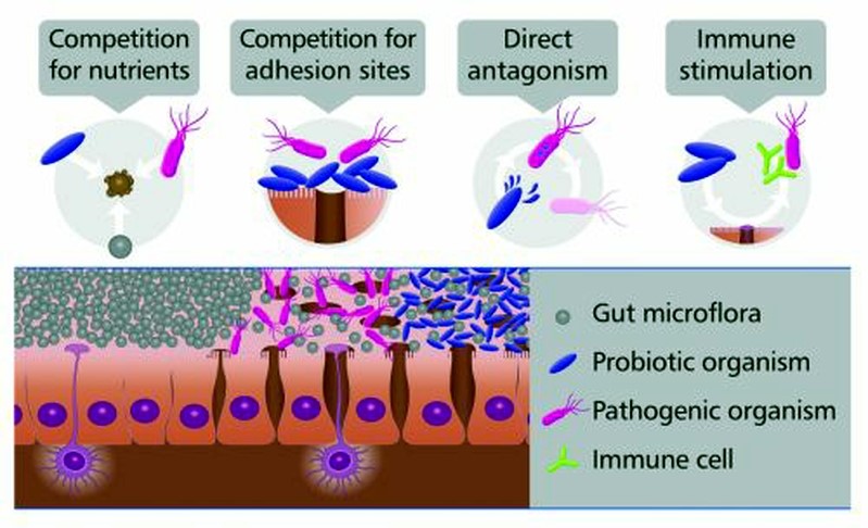
Figure 1 Depiction of the different ways in which the gut microbiome competes with invading cells, thereby inhibiting disease.
On the bottom, epithelial cells can be seen with the interior of the colon above them.
The purple cells with the long, extended appendages are immune cells.
Source.
Uneasy upheaval
With all these benefits, it is not surprising that an upsetting of the intestinal ecosystem of your dog can have very severe consequences for its health.
There are several ways in which the alteration of this ecosystem could affect dog health.
First of all, a (partial) collapse of this ecosystem may allow a pathogenic bacterium to invade, as alluded to before.
Secondly, a collapse or perturbation of this ecosystem may cause a bacterium that normally occurs only in low densities,
in which case it is harmless, to suddenly rise to ecological dominance, subsequently causing harm(5).
The latter mechanism is often implicated in the occurrence of acute and chronic diarrhoea (2,5,12,13)
Recently, researchers have successfully remediated several diseases that were caused by a similar collapse of the gut ecosystem in humans, through a procedure known as a faecal transplant.
In this procedure, the collapsed gut ecosystem of a patient is restored using faeces from a healthy individual (14).
However, to date, this has not been tried in dogs.
In the remainder of this article several of the species inhabiting the dog colon, and the faecal material contained inside it, will be examined in more detail.
Clostridium perfringens
Clostridium perfringens is one of the more evil inhabitants of the dog intestine.
The bacterium is rod-like in shape (a bacillus shape), see figure 2.
It is an obligate anaerobic organism, which can form endospores: extremely durable survival capsules.
When conditions are right, one spore gives rise to one bacterium, which can then reproduce normally.
C. perfringens is not only found in the dog, but also in soil and our own gut.
Its malignancy stems from the fact that it produces many toxins (15).
The bacterium causes food poisoning type A, the second most common bacterial infection in the USA (16).
Recently, it was also implicated in causing diarrhoea in a pet dog.
While this sounds evil alright, data shows us that Clostridium perfringens can also be present without inducing illness and diarrhoea (13).
This is in accordance with the view of the dog intestine as an ecosystem, in which a disbalance between the different inhabitants can rapidly lead to illness, as explained previously.
The question that arises is how Clostridium perfringens causes or is associated with diarrhoea. What do the toxins it produces do?
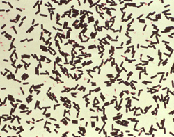
Figure 2 A culture of C. perfringens. The cells are clearly bacilli (rod-like in shape).
Source.
Clostridium toxins
Five types of Clostridium perfringens are recognised, based on the type of toxins they produce (15).
The most important toxin is called CPE, for Clostridium perfringens enterotoxin. The problem starts when vegetative (i.e. dormant, non-reproducing) C. perfringens are ingested.
Most of these will be annihilated by the acid bath in the stomach, but some survive.
These replicate, after which they begin forming endospores. During this sporulation, they produce CPE.
When the endospore is complete, the mother cell lyses, spilling its contents into the intestines.
This brings the CPE into the gut, where it can exert its effects (18).
Histopathological damage
The primary effect of the toxin is a histopathological one: it disrupts and damages the tissues lining the gut (18).
It causes upheaval in fluid and electrolyte flow,
and visible damage, e.g. shortening the intestinal villi and peeling off the upper epithelial layer.
The damage can be seen in figure 3. The left photo is of an intestinal model treated with vegetative C. perfringens, the right photo of a model treated with sporulating C. perfringens.
The villi that are numerous on the left photo are significantly reduced on the right photo, and many epithelial cells are lost (19).
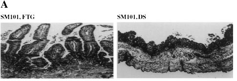
Figure 3 Model guts treated with either vegetative or sporulating C. perfringens. On the left, the lining is healthy, with many villi to enlarge the surface area of the intestines.
On the right, much of the epithelium has been killed off.
Source.
CPE's destructive effects
How is the destruction of these tissues brought about? CPE actually has two separate effects.
One is that it directly kill cells, the other is that it disrupts tight junctions
that help separate the gut from the rest of the body (see figure 4).
In direct killing, the CPE binds to certain receptors on the cell and forms a small complex with them on the cell membrane. The small complex is an important intermediate stage,
since it does not assemble on CPE-insensitive cells.
However, cells incubated at 4 degrees Celsius with CPE form the small complex, but do not perish.
It is only when the temperature is changed to 37 degrees Celsius that cell death sets in, which occurs concomitantly with the formation of larger CPE complexes.
The small complex interacts with other (unknown) molecules to produce the intermediate complex.
This is toxic because it forms pores in the cell membrane and the predominant form in cell cultures (16,18).
Normally, the cell membrane is semi-permeable, i.e. the substances that can pass through it are carefully regulated (6).
When pores occur in the cell membrane, this ability to regulate in- and outflow is disturbed. The outcome for the cell is disastrous and deadly (16,18).

Figure 4 Tight junctions make cells adhere so strongly to each other that leakage through them is nigh impossible.
This is achieved by proteins across the cell surface that have very strong interactions.
Source.
Tight junction destruction
What about the tight junctions (TJs)? In 1997, two receptors that interact with CPE were found.
Mouse cells that did not have these receptors were impervious to CPE damage.
When these cells were transformed so that they did possess these receptors, they responded to the toxin and perished.
This was the first clue in the search for the mechanism behind CPE TJ disruption (21).
The receptors in question are now known as claudin-3 and claudin-4. CPE kan bind to many members of the claudin protein family.
Another culprit was identified when it was shown that CPE can also bind to occludin,
but only when CPE itself has already bound to one of the claudins (22).
Claudins and occludin are important structural proteins in epithelial tight junctions (18,23).
Epithelial tight junctions keep cells closely tethered to one another.
The intestines are filled with food and bacteria, so it is of enormous importance that any uptake of nutrients is carefully controlled,
and that nothing can pass through unchecked (6).
CPE binds to proteins involved in forming these tight junctions, the claudins and occludins, thereby disrupting them.
While the primary effect is cell death in the (dog) colon, the subsequent disruption of the tight junctions worsens the leakage of fluids into the gut and thereby the diarrhoea.
Clostridium perfringens causes intestinal unease and diarrhoea in humans and dogs alike, and they are the source of the second largest bacterial infection in the USA in humans.
From the poop to the toxin that forms pores and disrupts tight junctions.
A remarkably complex mechanism, and just one of the myriad miracles that we stumble upon when inspecting dog faeces.
The Lactobacilli
What more can we learn about the dog gastrointestinal tract by investigating the steaming meadow muffin it bakes on a sunday morning?
Another collection of critters that can be found within the dog colon are bacteria of the genus Lactobacillus.
This genus currently contains 106 different species, some of which inhabit the dog colon (2,24).
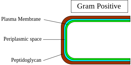
All Lactobacilli are gram positive, non-spore-forming bacteria that are slightly rounded in shape (coccobacilli, see figure 5).
These bacteria produce their energy by fermenting carbohydrates and producing lactic acid (24).
The presence of these bacteria is generally considered beneficial to the host's health (25).
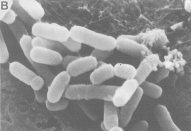
Figure 5 A scanning electron microscope image of Lactobacillus acidophilus, a species commonly used in probiotic products, such as yoghurt.
Adapted from this source.
Lactobacilli and host health
An important reason for this possible health benefit is related to the manner in which these bacteria obtain their energy.
The lactobacilli and many other gut bacteria obtain their energy through the fermentation of carbohydrates.
This fermentative process produces short-chain fatty acids.
These compounds provide extra energy to the host, aid the proliferation of the cells in the colon wall and are considered anticarcinogenic (27).
Alternatively, other bacteria in the colon obtain their energy through the fermentation of proteins, a process also known as putrefaction.
Although some short-chain fatty acids are produced during putrefaction, many toxic and possibly carcinogenic by-products are also created, such as ammonia, hydrogen sulphide and phenols.
A higher presence of lactobacilli seems to be able to shift the community composition in the colon towards bacteria that ferment carbohydrates and seems to
lower the occurrence of bacteria that obtain their energy through putrefaction (28).
Furthermore, lactobacilli seem to be able to counteract some of the detrimental effects caused by putrefying bacteria (27).
Several mechanisms have been proposed as to how lactobacilli manage to shift the composition of the faecal microbiome toward bacteria that ferment carbohydrates.
First of all, since they adhere tightly to the colon wall, they compete for space on the wall of the colon with other bacteria, including putrefying bacteria.
Secondly, lactobacilli are able to compete for nutrients, with putrefying bacteria. Thirdly, lactobacilli produce several compounds, such as bacteriocin and lactic acid, that inhibit the growth of certain other bacteria.
These three properties also make lactobacilli very effective at suppressing (opportunistic) bacterial pathogens such as C. perfringens (10,27,29,30)
This profound influence on the colonic microbial community is not the only health benefit provided by lactobacilli.
Since lactobacilli are gram-positive bacteria they possess a thick outer layer of the cell wall made of peptidoglycan(see figure 6).
This cell wall is surrounded by a polysaccharide structure, known as a capsule (6,27).
The polysaccharides in the capsule and the peptidoglycan in the cell wall are able to adsorb certain carcinogenic and mutagenic compounds.
Furthermore, the polysaccharides and peptidoglycan are able to adsorb primary bile acids. These compounds are essential to the digestion of fat and released into the small intestine.
The majority of these primary bile acids are resorbed at the end of the small intestine, however roughly 20% enter the colon.
In the colon, certain bacterial residents may convert these primary bile acids into secondary bile acids, which are mutagenic.
By adsorbing some of the primary bile acids entering the colon, the lactobacilli prevent the production of the potentially harmful secondary bile acids (27).
 As mentioned before, the faecal microbiome in the colon is also able to stimulate the immune system.
There are several ways in which lactobacilli stimulate the immune system, such as the stimulation of the mucosal barrier protecting the colon, the stabilisation of tight junctions and the reduction of inflammation.
Some of these effects may be induced by interactions between the lactobacilli and toll-like receptor 2.
This particular receptor is present on immune cells, such as the cells lining the colon and macrophages.
It is activated by binding to peptidoglycan and other components of the cell wall of gram-positive bacteria.
Experiments have shown that the activation of the toll-like receptor 2 in macrophages, amongst other effects, inhibits the production of pro-inflammatory cytokines.
Pro-inflammatory cytokines normally cause the initiation of an inflammatory response. By inhibiting the production of these compounds, lactobacilli may prevent inflammation in the colon (27).
As mentioned before, the faecal microbiome in the colon is also able to stimulate the immune system.
There are several ways in which lactobacilli stimulate the immune system, such as the stimulation of the mucosal barrier protecting the colon, the stabilisation of tight junctions and the reduction of inflammation.
Some of these effects may be induced by interactions between the lactobacilli and toll-like receptor 2.
This particular receptor is present on immune cells, such as the cells lining the colon and macrophages.
It is activated by binding to peptidoglycan and other components of the cell wall of gram-positive bacteria.
Experiments have shown that the activation of the toll-like receptor 2 in macrophages, amongst other effects, inhibits the production of pro-inflammatory cytokines.
Pro-inflammatory cytokines normally cause the initiation of an inflammatory response. By inhibiting the production of these compounds, lactobacilli may prevent inflammation in the colon (27).
Utilising Lactobacilli: pre-, pro- and synbiotics
The positive effects of lactobacilli mentioned above are nothing to shake a stick at.
It is not surprising, then, that researchers have tried to artificially increase their occurrence within the colon.
They have done so through the use of probiotics, prebiotics and synbiotics.
Probiotics are preparations of live lactobacilli or other beneficial bacteria that are simply ingested,
with the goal that some survive the acid bath in the stomach and will settle in the colon exerting their health benefits (29,30).
Prebiotics are substances, usually carbohydrates, that promote the growth of certain beneficial bacteria residing in the colon, such as lactobacilli, when added to the diet.
When prebiotics and probiotics are combined this is known as a symbiotic (27).
These three strategies have all been used in dogs, and there is some evidence that the use of synbiotics may provide the greatest health benefits (29,32).
Furthermore, probiotics are being used in humans on a massive scale, to which the numerous probiotic milk products in your local grocery store, many of which contain lactobacilli, bear witness.
Lactobacilli may be the true heroes of this gut-wrenching story on the dog gut faecal microbiome.
They can be very effective at suppressing microscopic bacterial pathogens, but they are helpless to prevent the occurrence of the harmful macroscopic critter inhabiting the dog colon that you are about to meet.
Toxocara canis
A very different poop inhabitant is Toxocara canis. Not a microbial member, but a eukaryotic parasite, and not a subject for the weak of stomach.
T. canis is a roundworm, a member of the genus Ascaris, that infects dogs and lives freely in their intestines, feeding off the food they eat.
The parasites are part of a whole range of protozoan and wormlike parasites that can infect dogs.
Live specimens can appear in the faeces or vomit of infected dogs, especially puppies (see figure 7), and can cause serious illnesses in them (33).
In addition, dogs shed the eggs in their faeces. However, these worms do not only pose a threat to our canine companions.
Indeed, humans can be infected by T. canis as well, and many urban areas are rife with their eggs.

Figure 7 T. canis in the faeces of a puppy. If left untreated, the puppy can suffer severe complications.
Source.
T. canis infection in puppies and bitches
The major source of infection for puppies are their mothers.
If an infection occurs sometime in the life of a bitch, the larvae of the worm penetrate the gut wall and travel into the tissues of the body.
These so-called somatic larvae can then lie dormant for years, only to be activated when the dog becomes pregnant.
They can then travel to the womb and infect the unborn puppies.
The infected puppies can expel worm eggs in their faeces in about two weeks after birth.
In addition, the larvae can be transferred via the mother’s milk, which means infection can take place until at least 38 days after birth.
The bitch does not emerge unscathed herself.
The larvae have lain dormant in the mother dog, but after being reactivated, they can often find their way back to her intestines and infect her, leading to T. canis eggs expelled in her faeces as well.
With each female worm producing up to 200,000 eggs per day, it quickly adds up.
It is simple to appreciate that these facts combined lead to easy propagation of a T. canis infection (33).
Symptoms in dogs
One of the problems in T. canis control is that mild infections are often not accompanied by symptoms.
When an infection becomes symptomatic, these symptoms are often coughing, frothy discharge from the nose, pneumonia and fluid accumulation in the lungs, all caused by larval migration.
Death can occur, especially in puppies that were infected within the mother.
Adult roundworms within puppies cause mucoid enteritis, an inflammation of the mucus lining the intestines, with symptoms ranging from vomiting to diarrhoea, anaemia (blood deficit), and emaciation.
Similar symptoms exist in older dogs, though they are often much less severe (33).
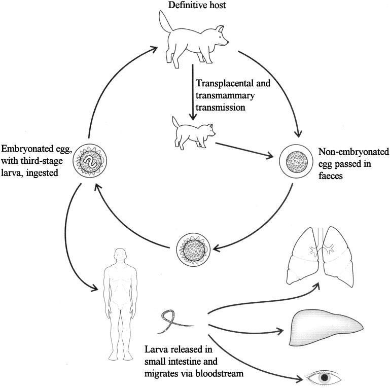
Figure 8 How dog infection and human infection are linked. Embryonated eggs can be picked up from the environment by humans. When ingested, complications can arise.
Source.
Risk for humans
What does this mean for us?
Infected dogs expel worm eggs in their faeces.
These eggs then survive in the urban parks and meadows where people walk their dogs (34).
Most of the roundworms carried by dogs are potentially infectious to humans as well.
In fact, T. canis is seen as a major cause of ascarid infection in humans, and toxocariasis, as it is called,
is the most prevalent worm infection in some countries (34).
The main mode of infection for humans is by contact with eggs in soil (see figure 8).
When the eggs are ingested, the larvae penetrate the intestinal wall and arrive in the bloodstream, and can spread through the body.
The larvae fail to turn into adults (since humans are not their main hosts), but they do cause local damage (33)
Two major syndromes may arise from roundworm infections in humans.
One is VLM, visceral larva migrans, the other is OLM, ocular larva migrans.
In VLM, the larvae infest the liver, lungs or the brain.
The symptoms include fever, rise in white blood cell levels, abdominal distress and pain, inflammation of the lungs with wheezing and even problems with the central nervous system.
In OLM, the activities of the larvae can lead to loss of sight(33).
Even though we are not the final host of T. canis, it can cause us plenty of discomfort.
End of tour
This will be the end of our tour of the dog colon and the excrement contained within it.
Right ahead of you, you’ll see the exit, on your left and right, the walls of the rectum.
No longer is dog excrement the repugnant stinking mess that it once was.
Well, to be honest, it still is, but its stink has gained a new dimension.
This intensely urban phenomenon harbours many interesting organisms.
We have learned that the faecal matter of the dog can be indicative of a whole ecosystem in its intestines.
We visited Clostridium perfringens, with its intricately complex, and still not completely resolved, mechanism of inducing illness.
We scooted past the Lactobacilli, shortly basking in their positive influence on the dog’s well-being, before treading in the murky mires of Toxicoma canis infection.
And what have we learned?
Dog faeces harbour an exquisite collection of life,
remnants of a fully functioning ecosystem that one seldom considers in daily life, an ecosystem that has members both benevolent and malicious towards the dog,
an ecosystem that plays host to parasites that can even have consequences for us.